What is
CARDIAC IMAGING?
Cardiac Imaging specializes in using advanced technologies to produce detailed images of the heart and its functions. As a highly innovative part of cardiology, the advent of new technologies such as three dimensional modeling to help in functional analysis of Cardiac Images has helped cardiologists gain a clearer insight into each patient's unique cardiovascular health.
Working as part of our Cardiology Hospital in Dubai, our cardiologists use these images are used to diagnose and monitor a variety of cardiovascular conditions, including coronary artery disease, heart failure, and valvular heart disease as well as blood flow.
Our principal patients include those with suspected or known cardiac conditions, as well as those preparing for cardiac surgery or other procedures.
MEET OUR CARDIOLOGY TEAM
- All
- Cardiology
Technologies used in Cardiovascular Imaging
Cardiovascular imaging modalities are varied, they include echocardiography, nuclear cardiology, MRI, and cardiovascular computed tomography (CT). Each of these technologies allows us to get a unique and detailed view of the heart and its functions.
Echocardiography
Echocardiography, also known as an echocardiogram or cardiac ultrasound, is a medical imaging test that uses high-frequency sound waves to produce images of the heart and its functions. It is a non-invasive and painless test that can be used to assess the heart's structure and function, as well as to monitor the effectiveness of treatments.
There are several different types of echocardiography, including transthoracic echocardiography, transesophageal echocardiography, and stress echocardiography. Transthoracic echocardiography is the most common type and involves placing a handheld device, called a transducer, on the patient's chest. The transducer sends out sound waves that bounce off the heart and produce echoes, which are then detected by the transducer and used to create images of the heart.
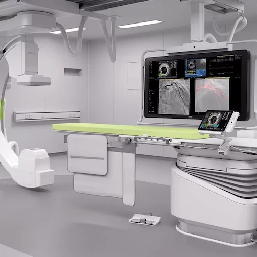
Transesophageal echocardiography involves inserting a small transducer into the patient's esophagus, which is located behind the heart. This allows for a clearer view of the heart and its functions.
Stress echocardiography involves performing an echocardiogram while the patient is exercising or after they have been given medication to increase their heart rate. This allows the cardiologist to assess how the heart functions during times of increased demand.
Echocardiography can be used to diagnose a variety of cardiovascular conditions, including coronary artery disease, heart failure, valvular heart disease, and abnormalities of the heart's structure. It can also be used to monitor the effectiveness of treatments, such as medications or procedures, and to plan for surgery.
Nuclear cardiology
Nuclear cardiology is a type of nuclear imaging that focuses specifically on the heart and its functions. It involves injecting a small amount of mildly radioactive material tracer into a vein and using a special camera to detect the tracer as it circulates through the heart and its major blood vessels.
Nuclear cardiology can be used to diagnose and monitor a variety of cardiovascular conditions, such as coronary artery disease, heart failure, and valvular heart disease.
The tracer is injected into a vein and travels to the heart, where it emits gamma rays that are detected by a special camera. This allows the cardiologist to see how well the heart is pumping and how much blood is flowing to different areas of the heart muscle.
There are several different types of radioactive material tracers that can be used in nuclear cardiology, including a radioactive substance called thallium, sestamibi, and tetrofosmin. Each radioactive material tracer has its own unique properties and can be used to assess different aspects of the heart's function.
Nuclear imaging in cardiology is typically used to assess the function of the heart and its major blood vessels, including the coronary arteries. It can be used to diagnose and monitor a variety of cardiovascular conditions, such as coronary artery disease, blood flow, heart failure, and valvular heart disease.
Positron emission tomography
Positron Emission Tomography or PET is a highly sensitive and specialized imaging modality that can be used to assess a wide variety of medical conditions, including cancer, neurological disorders, and cardiovascular diseases. It is particularly useful for detecting and monitoring changes in tissue function and metabolism, which can be important for diagnosis and treatment planning.
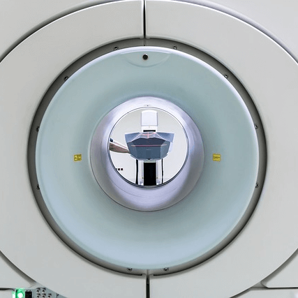
Magnetic Resonance Imaging (MRI)
MRI uses a powerful magnetic field and radio waves to produce detailed images of the body's structures and functions. MRI is a non-invasive and painless test that can be used to assess the heart's structure and function, as well as to detect abnormalities such as scar tissue or damaged areas of the heart muscle.
Computed tomography
Computed tomography (CT) scans are medical imaging tests that use x-rays and a computer to produce detailed cross-sectional images of the body's structures and functions.
CT scans are quick and painless and can be used to assess the heart's structure and function, as well as to detect abnormalities such as calcifications or blockages in the coronary arteries.
Cardiologists use CT scans in cardiac imaging to get detailed and accurate images of the heart and its surrounding structures, including the coronary arteries, valves, and major blood vessels.
CT scans can be used to diagnose and monitor a variety of cardiovascular conditions, such as artery disease, heart failure, and valvular heart disease. They can also be used to guide interventional procedures, such as stenting or angioplasty, and to assess the results of these procedures.
One of the main advantages of CT scans is their speed and convenience. CT scans can be performed quickly, often in just a few minutes, and do not require any special preparation, such as fasting or the use of contrast agents. This makes them a convenient and efficient option for patients who need to undergo cardiac imaging.
CT scans have the ability to produce detailed and accurate images of the heart and its surrounding structures. CT scans can detect a wide variety of abnormalities, including calcifications, blockages, and abnormalities in the heart's structure and function. They can also be used to assess the results of interventional procedures, such as stenting or angioplasty, and to monitor the effectiveness of treatments.
REQUEST AN APPOINTMENT
A member of our team will reach out to you & quickly get you booked in for an appointment with the most relevant member of our cardiology team.
PARTNERING WITH LEADING
MEDICAL INSURANCE PROVIDERS
We work with leading medical insurance providers in the country, if you have any questions or queries just give us a call on 8008254268.
